The new specimen, named *Pterygornis dapingfangensis*, belongs to the more advanced enantiornithines. Its anterior sternal margin develops a lateral sternal manubrium, a structure not reported in other Early Cretaceous birds, where the lateral sternal manubrium is primarily used for attachment to the clavicle membrane of the sternum.

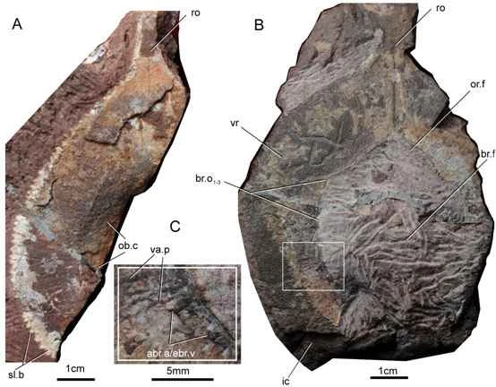
Figure 1. Holographic model and partial skull skeleton of the Great Flat-winged Winged Bird
Although the Early Cretaceous enantiornithine specimens are relatively complete, the overlapping of different bones makes it difficult to observe the morphological features of their skulls. The new specimens have dispersed skeletons, especially the smaller skull bones, which are well preserved, allowing for the reconstruction of some skull morphologies, particularly the zygomatic and quadrate zygomatic bones.
According to researchers, the zygomatic bone and quadrate zygomatic bone originate from different ossification centers. In the embryonic stage of modern birds, they are already completely fused into a rod-shaped bone, forming the lower edge of the eye socket. However, in dinosaurs, the close relatives of birds, the zygomatic bone and quadrate zygomatic bone do not fuse. The posterior end of the zygomatic bone bifurcates to form the postorbital process and the quadrate zygomatic process, while the quadrate zygomatic bone is T-shaped, extending anteriorly, dorsally, and posteriorly, with the zygomatic process, squamous process, and ventral posterior process extending outwards. In dinosaurs, the postorbital process of the zygomatic bone articulates with the postorbital bone, completely separating the eye socket from the inferior temporal fossa; the squamous process of the quadrate zygomatic bone articulates with the squamous bone, forming the closed posterior edge of the inferior temporal fossa. Compared to dinosaurs, the skulls of modern birds have undergone significant changes, especially the degeneration of the posterior part of the eye socket—the postorbital bone, and the loss of the zygomatic-postorbital joint and the quadrate zygomatic-squamous joint. These changes allow birds to use the movement of the quadrate bone in the anterior-posterior direction to push the palatal bones forward and backward, ultimately allowing the beak to rise or fall relative to the head for feeding activities.
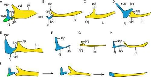
Figure 2. Evolution of the zygomatic bone and its morphology in dinosaurs and birds.
For a long time, due to the preservation of fossils, the details of how the aforementioned changes in the postorbital bones occurred during the evolution of dinosaurs to birds remained unclear. Through detailed comparison, Wang Min et al. discovered for the first time that the square zygomatic bone of winged birds is similar to that of Archaeopteryx, Confuciusornis, Huibird, Jeholornis, and Archaeopteryx, all having lost the ventral lateral process, resulting in an "L"-shaped skeleton. Compared to dinosaurs, the square zygomatic bone and zygomatic bone became slender in primitive birds, while the squamous and postorbital processes were significantly reduced. It is speculated that the joints between the zygomatic bone and the postorbital bone, and between the square zygomatic bone and the squamous bone, were already absent in birds of the Early Cretaceous. Furthermore, the "T"-shaped square zygomatic bone underwent an "L"-shaped transitional stage during its evolution towards a short-stalked form, and was the first to lose the ventral lateral process. This indicates that this skull feature, which is beneficial for birds' foraging activities, appeared in the early stages of bird evolution.